How To Read A Sonogram
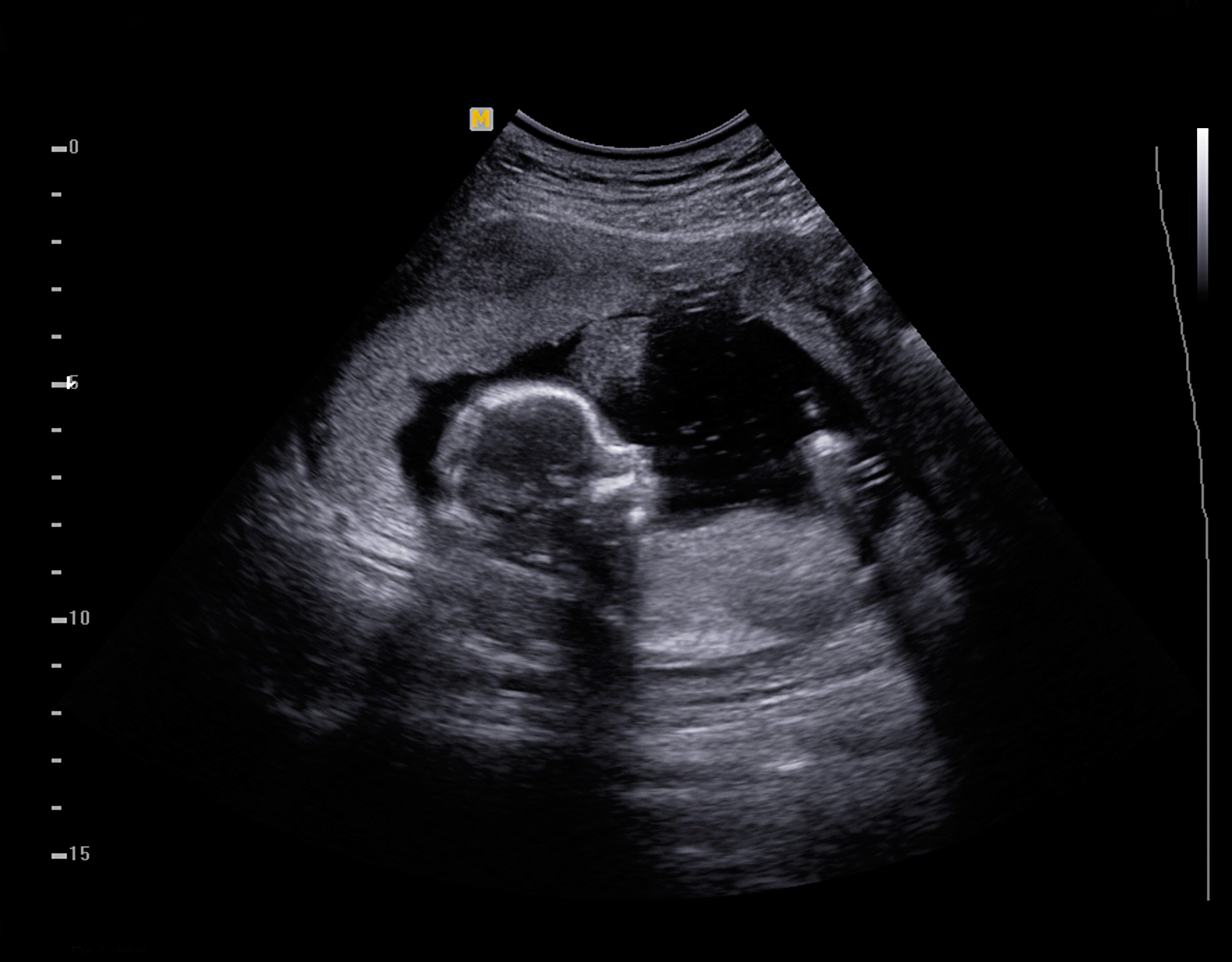
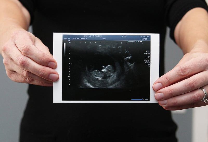
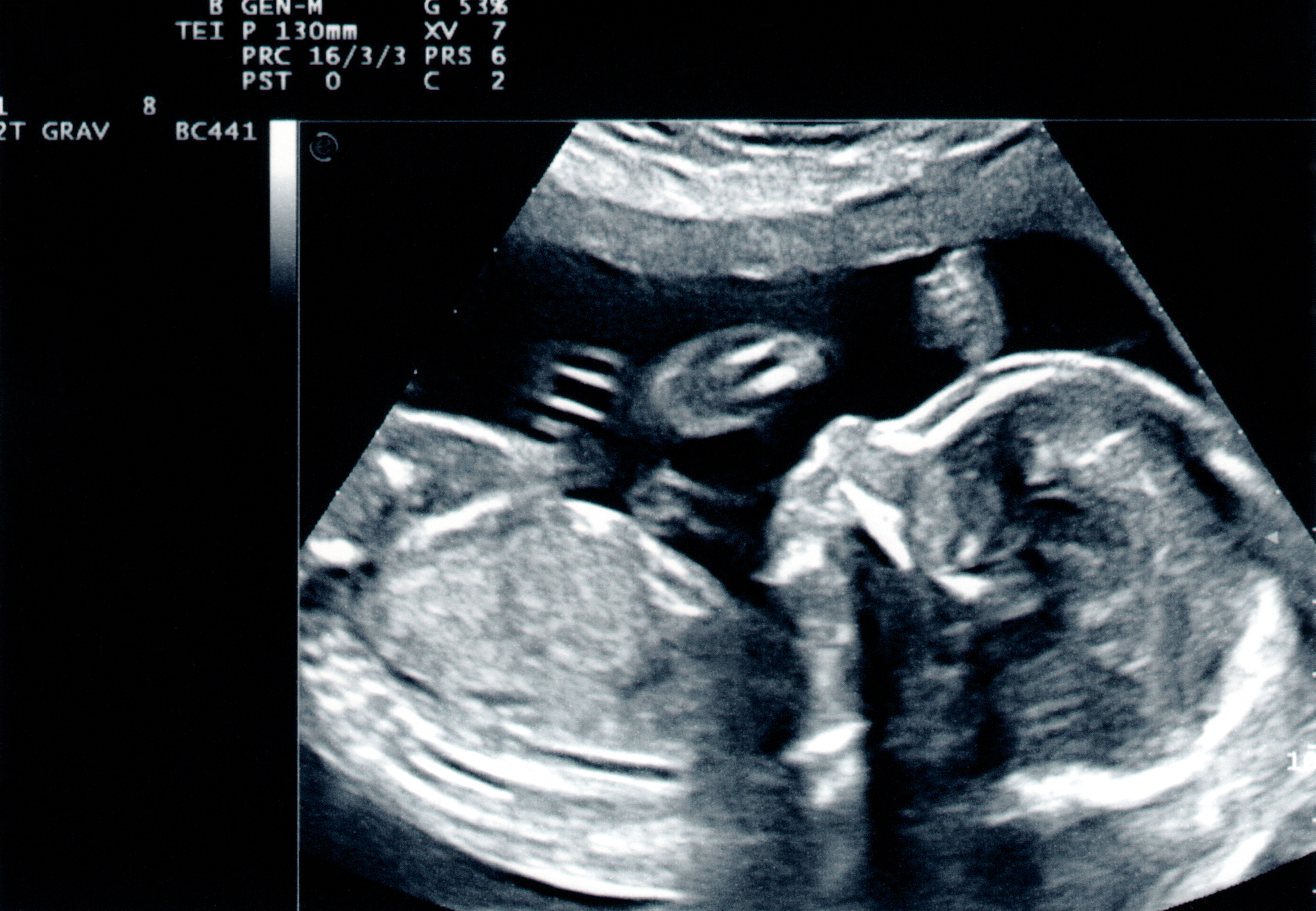
How To Read A Sonogram - The top of an ultrasound image usually shows a series of numbers and other information. Web sonogram definition, the visual image produced by reflected sound waves in a diagnostic ultrasound examination. Orientation you have to determine the orientation of. Sensors attached to the chest and sometimes the legs check the heart. The test is also called echocardiography or diagnostic cardiac ultrasound. Web during a fetal ultrasound, a device called a transducer is placed on the pregnant person's belly. Web the doctor or ultrasound technologist moves the transducer over the part of the body being studied. The computer inside the main part of the machine analyzes the signals and puts an image on the display screen. Web the global ultrasound device market is expected to experience significant growth, reaching a size of usd 14.5 billion by 2030, with a compound annual growth rate (cagr) of 4.3% during the forecast. Patient name hospital reference number ultrasound.
Web ultrasound (also called sonography or ultrasonography) is a noninvasive imaging test. Web how to read an ultrasound 1. Ultrasound centers and hospitals tend to use this space for details such as: This test checks the health of your abdominal organs — like your liver, gallbladder and kidneys — and the blood. Web sonogram definition, the visual image produced by reflected sound waves in a diagnostic ultrasound examination. Web however, the best way to define the contract between sonogram vs ultrasound would be this: Or in some cases, it may be placed in the vagina or on the area between the vagina and the anus. You can also see ultrasound numbers when obtaining a fetal image, aside from the image itself. Web explaining the ultrasound numbers. The computer inside the main part of the machine analyzes the signals and puts an image on the display screen.
Web the doctor or ultrasound technologist moves the transducer over the part of the body being studied. Here’s a brief explanation of how to read ultrasound numbers and what they mean: They are trained in interpreting and analyzing different medical images. Sonography is the use of an ultrasound. The top of an ultrasound image usually shows a series of numbers and other information. The ultrasound is the process to retrieve the information and the sonogram is the end picture showing the result. A sonogram is the picture that the ultrasound generates. Web ultrasound (also called sonography or ultrasonography) is a noninvasive imaging test. Web however, there’s a difference between the two: The test is also called echocardiography or diagnostic cardiac ultrasound.

Sonogram showing baby giving thumbs up in the womb goes viral ABC11
An ultrasound picture is called a sonogram. This appears as a hypo echoic pattern posterior to highly. An echocardiogram uses sound waves to show how blood flows through the heart and heart valves. Abdominal pain abnormal blood tests (often for blood tests. The test is also called echocardiography or diagnostic cardiac ultrasound.

estatenygw pregnancy week 6 ultrasound photos
Sonography is the use of an ultrasound. This test checks the health of your abdominal organs — like your liver, gallbladder and kidneys — and the blood. Web ultrasound (also called sonography or ultrasonography) is a noninvasive imaging test. Orientation you have to determine the orientation of. The test is also called echocardiography or diagnostic cardiac ultrasound.

Baby sonogram ornament please read full description before Etsy
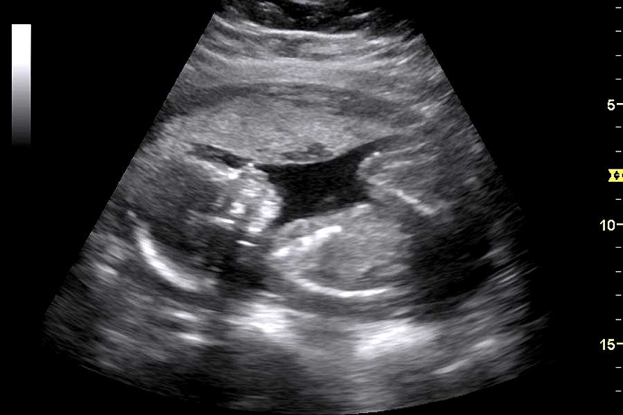
Orientation you have to determine the orientation of. Web the doctor or ultrasound technologist moves the transducer over the part of the body being studied. The top of an ultrasound image usually shows a series of numbers and other information. A sonogram is the picture that the ultrasound generates. Typically, the radiologist sends the report to the person who ordered.
First Look at Your Baby The Fascinating History of the "Sonogram"
Or in some cases, it may be placed in the vagina or on the area between the vagina and the anus. Web a baby sonogram, or fetal ultrasound, is an image to check the sex, motion, or development of a growing fetus. Web how to read an ultrasound 1. Web explaining the ultrasound numbers. This is the lack of sound.

baby sonogram YouTube
A sonogram is the picture that the ultrasound generates. Web what do the numbers mean at the top of an ultrasound image? Color an ultrasound or sonogram picture is a black and white photograph, so they all look the same to someone who. Abdominal pain abnormal blood tests (often for blood tests. The ultrasound is the process to retrieve the.
How to Read an Ultrasound Gender and And Abnormality? New Health Advisor
Patient name hospital reference number ultrasound. Web what do the numbers mean at the top of an ultrasound image? It generally indicates a solid internal consistency. The ultrasound is the process to retrieve the information and the sonogram is the end picture showing the result. Web how to read an ultrasound 1.
Sonogram vs Ultrasound A More InDepth Distinction Between The Two
However, it is important for health care providers to have a basic understanding of what an ultrasound. This appears as a hypo echoic pattern posterior to highly. Web explaining the ultrasound numbers. The radiologist writes the report for your provider who ordered the exam. Web spleen abdominal aorta and other blood vessels of the abdomen doctors use ultrasound to help.

6 Ways to Tell Baby's Gender From an Early Sonogram
An ultrasound reading is usually performed by the radiologist. Web an echocardiogram (echo) uses high frequency sound waves (ultrasound) to make pictures of your heart. Color an ultrasound or sonogram picture is a black and white photograph, so they all look the same to someone who. However, it is important for health care providers to have a basic understanding of.
OneCall24
This test checks the health of your abdominal organs — like your liver, gallbladder and kidneys — and the blood. Patient name hospital reference number ultrasound. Web however, there’s a difference between the two: Orientation you have to determine the orientation of. However, it is important for health care providers to have a basic understanding of what an ultrasound.
Sonogram SG1 free sonogram by agworks
Web however, the best way to define the contract between sonogram vs ultrasound would be this: Web what do the numbers mean at the top of an ultrasound image? Sonography is the use of an ultrasound. Patient name hospital reference number ultrasound. The top of an ultrasound image usually shows a series of numbers and other information.
The Radiologist Writes The Report For Your Provider Who Ordered The Exam.
Orientation you have to determine the orientation of. They are trained in interpreting and analyzing different medical images. Typically, the radiologist sends the report to the person who ordered your test, who then delivers the results to. An echocardiogram uses sound waves to show how blood flows through the heart and heart valves.
Web Explaining The Ultrasound Numbers.
You can also see ultrasound numbers when obtaining a fetal image, aside from the image itself. Web however, the best way to define the contract between sonogram vs ultrasound would be this: Web however, there’s a difference between the two: Sensors attached to the chest and sometimes the legs check the heart.
Web A Baby Sonogram, Or Fetal Ultrasound, Is An Image To Check The Sex, Motion, Or Development Of A Growing Fetus.
Web how to read an ultrasound 1. The computer inside the main part of the machine analyzes the signals and puts an image on the display screen. Web an echocardiogram (echo) uses high frequency sound waves (ultrasound) to make pictures of your heart. An ultrasound picture is called a sonogram.
Abdominal Pain Abnormal Blood Tests (Often For Blood Tests.
This appears as a hypo echoic pattern posterior to highly. Web the doctor or ultrasound technologist moves the transducer over the part of the body being studied. A sonogram is the picture that the ultrasound generates. Color an ultrasound or sonogram picture is a black and white photograph, so they all look the same to someone who.